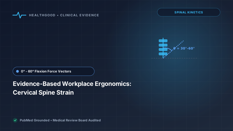
- Cervical Gravitational Load: Flexing the neck forward by just 45° increases the effective mechanical load on the cervical spine from 10–12 lbs to nearly 49 lbs (22 kg).
- Upper Crossed Syndrome (UCS): Chronic desk slouching precipitates reciprocal muscular imbalance: hypertonic upper trapezius and pectoralis muscles counterbalanced by inhibited deep cervical flexors and rhomboids.
- Monitor Sightline Alignment: The top third of the visual display terminal should align directly with horizontal eye level, maintaining a downward visual angle of 15° to 20°.
- Active Micro-breaks: Static postural loading exceeding 30 continuous minutes reduces muscular blood perfusion by 40%, necessitating brief dynamic 60-second postural resets.
Prolonged static computer use has emerged as the leading etiology of non-traumatic musculoskeletal disorders in modern sedentary workforces across the United States, United Kingdom, and Canada. Termed "Forward Head Posture" (FHP) or colloquial "tech neck," sustained forward translation of the cranium alters normal cervical lordosis and places enormous compressive stress across the C5–C7 intervertebral discs.
In a healthy cervical spine with neutral alignment, the gravitational center of the adult skull (weighing approximately 10 to 12 pounds) sits directly over the vertical axis of the thoracic vertebral bodies. As the head tilts forward, the physical lever arm lengthens, creating a disproportionate mechanical bending moment that requires constant isometric contraction from the posterior cervical extensor musculature.
Biomechanical Loading: Cervical Flexion vs. Effective Force
In a landmark finite-element biomechanical modeling study by Dr. Kenneth K. Hansraj, Chief of Spine Surgery at New York Spine Surgery & Rehabilitation Medicine (2014), the effective gravitational load exerted on the cervical spine was calculated across varying degrees of sagittal neck flexion:
| Flexion Angle (Degrees) | Effective Load (US Imperial lbs) | Effective Load (Metric kg) | Biomechanical Tissue Impact |
|---|---|---|---|
| 0° (Neutral Anatomical) | 10 – 12 lbs | 4.5 – 5.5 kg | Normal compressive stress distributed across facet joints |
| 15° (Slight Downward Tilt) | 27 lbs | 12.2 kg | Early trapezius and levator scapulae myofascial strain |
| 30° (Standard Laptop Screen) | 40 lbs | 18.1 kg | Substantial shear stress on C5-C6 discs; posterior ligament tension |
| 45° (Typing / Mobile Device) | 49 lbs | 22.2 kg | Progressive disc degeneration; loss of cervical lordotic curvature |
| 60° (Slumped Lap Viewing) | 60 lbs | 27.2 kg | Massive shearing force equal to carrying an 8-year-old child on neck |
Pathophysiology of Upper Crossed Syndrome (UCS)
Described originally by the Czech physiatrist Vladimir Janda, Upper Crossed Syndrome reflects a systematic neurological pattern of muscle facilitation (hypertonicity/shortening) and inhibition (hypotonicity/lengthening) across the cervicothoracic girdle:
- Facilitated (Shortened & Overactive): Pectoralis major and minor, upper trapezius, levator scapulae, and suboccipital muscles.
- Inhibited (Weakened & Hypotonic): Deep cervical neck flexors (longus colli, longus capitis), lower trapezius, serratus anterior, and rhomboids.
This imbalance forces the scapulae into anterior tilt and internal rotation (protracted, rounded shoulders) while hyperextending the occiput on the atlas (C1), frequently compressing the greater occipital nerve and generating chronic cervicogenic tension headaches.
The 90-90-90 Clinical Desk Setup Protocol
To neutralize gravitational shearing forces, the workplace workstation must be calibrated to the individual user's anthropometric dimensions following the evidence-based 90-90-90 orthopedic paradigm:
- Lower Extremities: Feet resting flat on the floor or on a rigid footrest, with knees bent at an angle of 90° to 100°. Seat pan depth should leave a 2-inch gap between the front cushion edge and the popliteal fossa (back of knee) to avoid compressing the sciatic nerve.
- Pelvis and Lumbar Spine: Hips flexed at 90° to 100° with the pelvis firmly anchored against the lumbar support of the chair. Maintaining 20° to 30° of lumbar lordosis naturally induces thoracic uprightness and eliminates compensatory forward craning of the neck.
- Upper Extremities: Armrests adjusted so shoulders remain relaxed (no shrugging) with elbows flexed at 90°. Forearms should run parallel to the floor, allowing wrists to remain in neutral alignment (0° to 15° extension) during typing, preventing median nerve compression within the carpal tunnel.
Monitor Distance and Sightline Placement
Display positioning is the primary visual driver of cervical posture. Follow these clinical parameters:
- Viewing Distance: Position the screen approximately one arm's length away (20 to 28 inches / 50 to 70 cm). Users should be able to touch the center of the display with outstretched fingertips.
- Vertical Height: The top third of the monitor casing should sit exactly at horizontal eye level. Because the human eye naturally rests at a downward declination of 15° to 20°, this configuration maintains neutral cervical balance without requiring neck flexion.
- Dual Monitor Symmetry: If dual monitors are utilized equally, angle them in an inward semi-circle with the center seam directly in front of the nose to prevent continuous unilateral sternocleidomastoid rotation.
Red-Flag Neurological Symptoms: Neck discomfort accompanied by radiating tingling, sharp burning down the arm, reduced hand grip strength, or sudden clumsiness dropping objects (cervical radiculopathy or myelopathy) requires prompt clinical imaging (MRI) and spine specialist consultation.
Evidence-Based Clinical Rehab: The 20-20-20 & Chin Tuck Regimen
Static isometric contraction impairs capillary blood flow. Implement this two-exercise physical therapy routine every 60 minutes:
- Cervical Retraction ("Chin Tucks"): Sit upright with shoulders pinned back. Without tilting the head up or down, retract the chin horizontally backwards as if making a "double chin." Hold for 5 seconds; repeat for 10 repetitions. This selectively strengthens the deep longus colli flexors.
- Thoracic Extension & Scapular Squeeze: Interlock fingers behind the occiput, open elbows wide, and gently arch the mid-back over the chair backrest while squeezing the shoulder blades together for 10 seconds.
Peer-Reviewed Scientific References
- Hansraj KK. "Assessment of stresses in the cervical spine caused by posture and position of the head." Surg Technol Int. 2014;25:277-279. PMID: 25393825
- Ariëns GA, Bongers PM, Douwes M, et al. "Are neck flexion, neck rotation, and sitting at work associated with neck pain? Results of a prospective cohort study." Pain. 2001;93(2):125-136. PMID: 11440737
- Page P, Frank CC, Lardner R. Assessment and Treatment of Muscle Imbalance: The Janda Approach. Human Kinetics; 2010.
- Cagnie B, Danneels L, Van Tiggelen D, De Loose V, Cambier D. "Individual and work related risk factors for neck pain among office workers: a cross sectional study." Eur Spine J. 2007;16(5):679-686. PMID: 17160393
- Neumann DA. Kinesiology of the Musculoskeletal System: Foundations for Rehabilitation. 3rd ed. Elsevier; 2017.